US-DATAData Annotation
Cabinet
We prepare medical images, studies and documents for AI training — organ and pathology segmentation, classification, detection, DICOM annotation and medical text.
From a test batch to a structured dataset for model training and validation.
Calculate project cost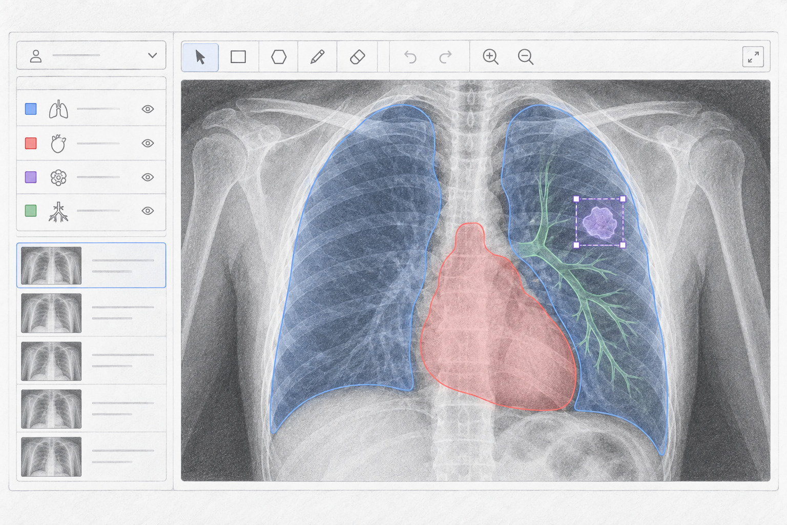
Medical data annotation is the preparation of images, studies, text and documents for training artificial intelligence models.
Depending on the task, annotators mark anatomical structures, organs, regions of interest and pathological changes, assign classes and label medical text.
Medical AI can use X-rays, CT, MRI, ultrasound, digital pathology, microscopy and other medical data types.
These projects are complex and require strict annotation consistency. Small differences in how the same region is outlined can change the structure of the final dataset.
Before scaling it is important to define classes, annotation criteria, the output format and rules for ambiguous cases.
Precise outlines of:
Regions of interest such as lesions, foci, damage, inflammatory areas and other changes defined by the specification. The format can include bounding boxes, polygons and segmentation masks.
Categories for an image or a study:
Bounding boxes or other outlines for individual objects and regions of interest.
Key anatomical landmarks and characteristic points.
Reports, protocols, clinical notes, entities and attributes. Information from forms, certificates, tables and other medical documents.
Different annotation types for computer vision tasks
Full data preparation cycle from raw data to model-ready output

How we keep medical annotation consistent
Medical data often contains complex structures, small regions of interest and ambiguous cases, so consistency requirements are especially high.
Before launch we fix classes, object rules and boundary requirements.
Multi-step review runs throughout the project, and disputed cases follow a pre-agreed logic.
Medical AI often works with specialized studies rather than ordinary photos.
US-DATA can organize annotation for tasks related to:
Depending on the specification we can mark organs, individual structures, regions of interest and pathological changes.
For image series it is especially important to keep the same rules across slices and studies.
The output format is agreed for the customer's ML pipeline and software.
Many Medical AI tasks need more than an approximate bounding box. The boundary of the target region must be defined precisely.
Segmentation creates pixel masks for:
These projects especially need shared boundary criteria and control of disagreement between annotations.
US-DATA organizes the process so the rules stay the same across the dataset.
Digital microscopy and histology images can contain a large number of small objects and regions of interest.
Depending on the task we can annotate:
Bounding boxes, polygons, point annotation, segmentation and classification can be used.
Data with a complex structure, small objects and ambiguous boundaries.
We follow the customer's instructions or help formalize the rules.
The output format can be adapted to the customer's ML pipeline.
We check accuracy, completeness and consistency.
We start with a test sample and scale after quality is agreed.
Annotation can run in the customer's system or in agreed software.
Consistent medical image annotation
Precise segmentation of regions of interest
A structured dataset for model training
Shared rules for hard and borderline cases
Data ready for training, testing and validation
Medical data requires extra attention to security and confidentiality. Only specialists assigned to the specific project receive access. If needed, annotation can run in the customer's infrastructure or software.
Expandable sections with indicative cost tables.
Choose parameters and get instant estimate
* This estimate is not a public offer. Final cost is determined after technical analysis and data review.
Latest materials on data annotation and machine learning
Share a data sample and describe the task — we will estimate the volume, annotation requirements and an approach to the dataset.
Artificial intelligence is widely used to analyze medical images, process documents and support research. These solutions need carefully prepared datasets with consistent annotation.
US-DATA provides image annotation and other data labeling for Medical AI. Depending on the task we use bounding boxes, polygons, segmentation masks, classification, keypoints and text annotation.
A key direction is CT, MRI and X-ray annotation. These studies can be labeled for anatomical structures, organs and specified regions of interest. Where object geometry matters, we use image segmentation. Individual objects and zones can also be marked with object detection.
Digital pathology and microscopy are a separate category. These images can contain many small structures, so the process needs detailed instructions and stable quality control.
US-DATA can also work with medical documents and text. That includes OCR, document classification, entity extraction and structured data for NLP, based on text data annotation.
Confidentiality and controlled access matter especially for medical projects. The process can follow the customer's requirements for infrastructure, file transfer and access separation.
Before scaling we recommend a test batch. It aligns classes, rules for hard cases and the final data format.
If your project needs medical image annotation, CT or MRI segmentation, Medical AI data, digital pathology or medical NLP, US-DATA will organize the process and prepare a dataset in the agreed format.